Additional Information
Book Details
Abstract
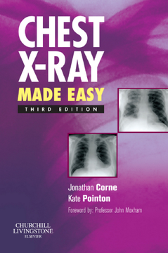
Translated into over a dozen languages, this book has been widely praised for making interpretation of the chest X-ray as simple as possible. It describes the range of conditions likely to be encountered on the wards and guides the doctor through the process of examining and interpreting the film based on the appearance of the abnormality shown. It then assists the doctor in determining the nature of the abnormality and points the clinician towards a possible differential diagnosis. It covers the common radiological problems the junior doctors are faced with starting with the appearance of the film, e.g. showing generalised shadowing or a coin lesion. It gives advice on how to examine an X-ray, how to check its technical quality and how to identify where the lesion is. All the X-rays are accompanied by a simple line diagram outlining where the abnormality is.
- Covers the full range of common radiological problems.
- Includes valuable advice on how to examine an X-ray.
- Assists the doctor in determining the nature of the abnormality.
- Points the clinician towards a possible differential diagnosis.
- Now presented in two-colour to enhance the appearance of the text.
- New material includes an introduction to thoracic CT scanning indicating the usefulness of these scans where appropriate.
Table of Contents
| Section Title | Page | Action | Price |
|---|---|---|---|
| Front cover | cover | ||
| half title page\r | i | ||
| Chest X-Ray Made Easy | iii | ||
| copyright page | iv | ||
| Foreword | v | ||
| Preface | vii | ||
| Acknowledgements | ix | ||
| Table of contens | xi | ||
| CHAPTER 1 How to look at a chest X-ray | 1 | ||
| 1.1 Basic interpretation is easy | 2 | ||
| 1.2 Technical quality | 4 | ||
| Projection | 8 | ||
| Orientation | 8 | ||
| Rotation | 8 | ||
| Penetration | 9 | ||
| Degree of inspiration | 9 | ||
| 1.3 Scanning the PA film | 10 | ||
| 1.4 How to look at the lateral film | 13 | ||
| CHAPTER 2 Localizing lesions | 17 | ||
| 2.1 The lungs | 18 | ||
| 2.2 The heart | 21 | ||
| CHAPTER 3 The CT scan | 27 | ||
| CT scanning | 28 | ||
| Types of CT scan | 28 | ||
| High-resolution CT scanning (HRCT) | 28 | ||
| Spiral CT | 29 | ||
| Combined imaging | 29 | ||
| Interpreting the images | 29 | ||
| Finding your way around the CT scan | 30 | ||
| Artefacts | 40 | ||
| CHAPTER 4 The white lung field | 41 | ||
| 4.1 Collapse | 42 | ||
| 4.2 Volume loss | 54 | ||
| 4.3 Consolidation | 58 | ||
| 4.4 Pneumocystis carinii (jiroveci) pneumonia (PCP) | 62 | ||
| 4.5 Pleural effusion | 64 | ||
| 4.6 Asbestos plaques | 68 | ||
| 4.7 Mesothelioma | 70 | ||
| 4.8 Pleural disease on a CT scan | 72 | ||
| 4.9 Lung nodule | 74 | ||
| 4.10 Cavitating lung lesion | 78 | ||
| 4.11 Left ventricular failure (LVF) | 82 | ||
| 4.12 Acute respiratory distress syndrome | 86 | ||
| 4.13 Bronchiectasis | 90 | ||
| The HRCT scan and bronchiectasis | 91 | ||
| 4.14 Fibrosis | 94 | ||
| The HRCT scan and pulmonary fibrosis | 95 | ||
| Confirming fibrosis | 96 | ||
| Determining aetiology | 97 | ||
| 4.15 Chickenpox pneumonia | 100 | ||
| 4.16 Miliary shadowing | 102 | ||
| CHAPTER 5 The black lung field | 105 | ||
| 5.1 Chronic obstructive pulmonary disease (COPD) | 106 | ||
| 5.2 Pneumothorax | 110 | ||
| 5.3 Tension pneumothorax | 112 | ||
| 5.4 Pulmonary embolus (PE) | 114 | ||
| scanning | 114 | ||
| CT pulmonary angiogram | 116 | ||
| 5.5 Mastectomy | 119 | ||
| CHAPTER 6 The abnormal hilum | 121 | ||
| 6.1 Unilateral hilar enlargement | 122 | ||
| 6.2 Bilateral hilar enlargement | 126 | ||
| CHAPTER 7 The abnormal heart shadow | 129 | ||
| 7.1 Atrial septal defect (ASD) | 130 | ||
| 7.2 Mitral stenosis | 132 | ||
| 7.3 Left ventricular aneurysm | 134 | ||
| 7.4 Pericardial effusion | 136 | ||
| CHAPTER 8 The widened mediastinum | 139 | ||
| CHAPTER 9 Abnormal ribs | 143 | ||
| 9.1 Rib fractures | 144 | ||
| 9.2 Metastatic deposits | 146 | ||
| CHAPTER 10 Abnormal soft tissues | 149 | ||
| Surgical emphysema | 150 | ||
| CHAPTER 11 The hidden abnormality | 153 | ||
| 11.1 Pancoast’s tumour | 154 | ||
| 11.2 Hiatus hernia | 156 | ||
| 11.3 Air under the diaphragm | 158 | ||
| Index | 161 | ||
| A | 161 | ||
| B | 162 | ||
| C | 162 | ||
| D | 163 | ||
| E | 163 | ||
| F | 164 | ||
| G | 164 | ||
| H | 164 | ||
| I | 165 | ||
| K | 165 | ||
| L | 165 | ||
| M | 166 | ||
| N | 166 | ||
| O | 167 | ||
| P | 167 | ||
| R | 168 | ||
| S | 168 | ||
| T | 168 | ||
| U | 169 | ||
| V | 169 | ||
| W | 169 |
